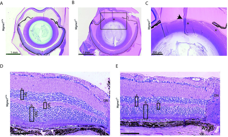
Fig 3. Mgme1-/- eye tissue histology images show clear alterations of lens and retina morphology.
(A) Sections through the Mgme1+/+ and (B) the Mgme1-/- eye, indicate at first glance alterations of the lens matrix structure (*); (C) enlarged Mgme1-/- anterior lens image reveals alterations of the lens fiber cells: they are swollen and disorganized (*), affecting the stability of the whole lens matrix. Additionally changes are present as to the epithelial cell monolayer, located underneath the lens capsule, shown here as an accumulation of epithelial cells that seem to make place for invaginations of the lens capsular material (arrow). (D) Retinal layers near ON images show an arbitrary thickness evaluation of some of the layers as rectangles (rectangle a—INL, rectangle b—ONL, rectangle c—OPL) in their ‘control’ size that in E. are placed over the Mgme1-/- retinal layers, to indicate the reduction in all three layers. Abbreviations: INL—inner nuclear layer, ONL—outer nuclear layer, OPL—outer plexiform layer, ON—optic nerve.

