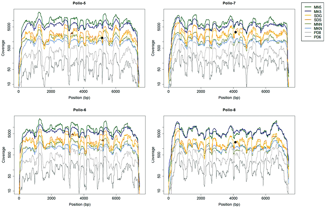
Fig. 5. Coverage patterns across the poliovirus genome.

The depth of coverage, plotted on a log scale, across the length of the genome is depicted for all datasets (denoted by color). Polio-5 and Polio-6 are both type 1 polioviruses, while Polio-7 and Polio-8 are type 3 viruses. Orange triangles indicate the positions of high frequency indels in the SDS consensus genome sequences, while black points indicate the positions of high-frequency indels found at the same position for both SDG and SDS datasets (only one point per position is shown for simplicity).
