
FIGURE 1.
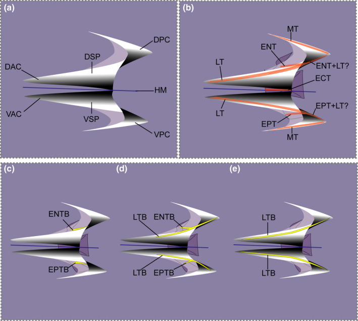
Schematic representation of typical myoseptum from midbody in lateral view (anterior to the left). (a) Position of cones in epaxial part (DAC, DPC) and hypaxial part (VAC, VPC) are shown. (b) Arrangement of possible tendons of a myoseptum is shown: epineural tendon (ENT), epipleural tendon (EPT), lateral tendon (LT), epicentral (ECT) and myorhabdoid tendon (MT). It appears as if the fibre bundles of the epineural and lateral tendons merge posteriorly (ENT+LT?/EPT+LT?). (c–e) Three possible ossification patterns resulting in an epineural bone/epipleural bone. (c) Ossification in the epineural tendon (ENTB)/epipelural tendon (EPTB) usually attaching to the neural arch/parapophysis, respectively. (d) Proximally forked epineural/epipleural bone representing ossifications in the epineural tendon/epipleural tendon (ENTB/EPTB) and the lateral tendon (LTB). (e) In myosepta further posteriorly the simple epineural/epipleural bone can be represented by an ossification in the lateral tendon only (LTB). Colour code: orange lines indicate tendons; yellow, ossified intermuscular bone; blue, horizontal midline