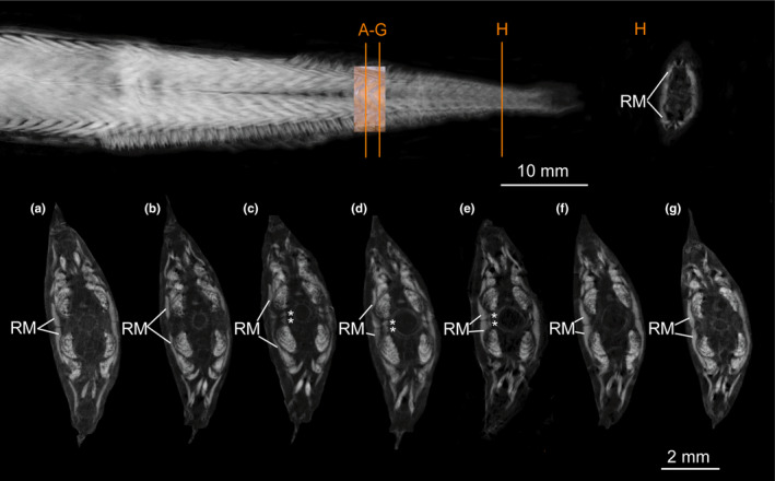
FIGURE 11.

Micro‐CT scan of Eustomias schmidti (USNM 261301, 185 mm SL) stained with iodine to visualize muscles. (a–g) Different sections of red musculature (RM) between middle part of a myomere to middle part of following myomere (orange lines in overview show positions of sections shown in (a) and (g). (a) Section through middle part of superficial RM. (b) Superficial RM spread dorsally and ventrally. (c–e) Paired bundles of RM appear (marked with an *) between superficial RM (of following myomere) and vertebral column and occupy space between widely separate dorsal and ventral anterior cones. (f–g) Middle part of following superficial RM (in these sections the paired bundles are not visible as they are tendinous (RMT). (h) Amount of superficial RM further posterior increased
