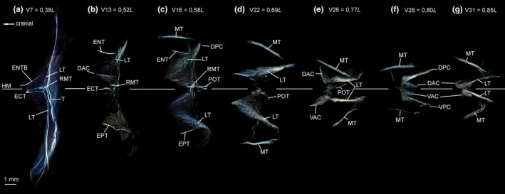
FIGURE 19.

Dissected myosepta of Argyropelecus affinis (MNHN 2021‐0224, 59 mm SL) photographed under polarized light. (a) Myoseptum of V7 at 0,38L. ECT, LTs and ENTB present. Posterior border of myoseptum tendinous (T). (b) Myoseptum of V13 at 0,52L. Anterior cones present. ECT, LTs, ENT and EPT present. (c) Myoseptum of V16 at 0,58L. As in (b) POT illustrated. (d) Myoseptum of V22 at 0,69L. LTs extending from anterior into posterior cones. (e) Myoseptum of V26 at 0,77L. Anterior cones elongated and turned dorsally/ventrally. (f) Myoseptum of V28 at 0,80L. DAC/VAC completely turned posteriad. (g) Myoseptum of V31 at 0,85L. DAC/VAC still turned posteriad, but not as much as in previous myosepta. Abbreviations: DAC, dorsal anterior cone; DPC, dorsal posterior cone; ECT, epicentral tendon; ENT, epineural tendon; ENTB, epineural tendon bone; EPT, epipleural tendon; HM, horizontal midline; LT, lateral tendon; MT, myorhabdoid tendon; POT, posterior oblique tendon; RMT, tendon within red muscle bundles; T, tendon at the posterior border of the myoseptum; VAC, ventral anterior cone; VPC, ventral posterior cone
