
Figure 2. Development of atherosclerosis lesions.
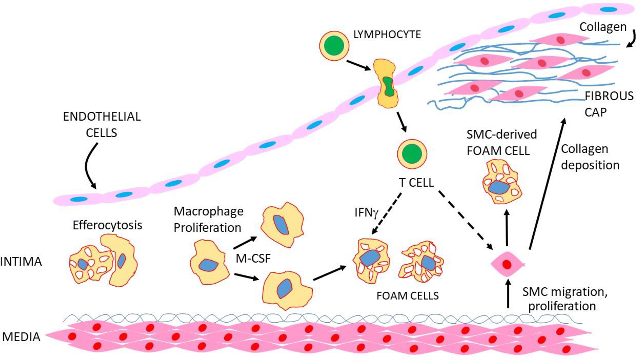
Macrophages proliferate in response to M-CSF, and foam cells are engulfed by macrophages, a process known as efferocytosis. SMC transform to a proliferative state, migrate to the endothelial region and secrete collagen to give rise to a “fibrous cap”. SMC can also transform to macrophage-like cells that take up lipid to give rise to foam cells. T and B cells also enter the lesion and interact with other cell types to promote or retard lesion development.