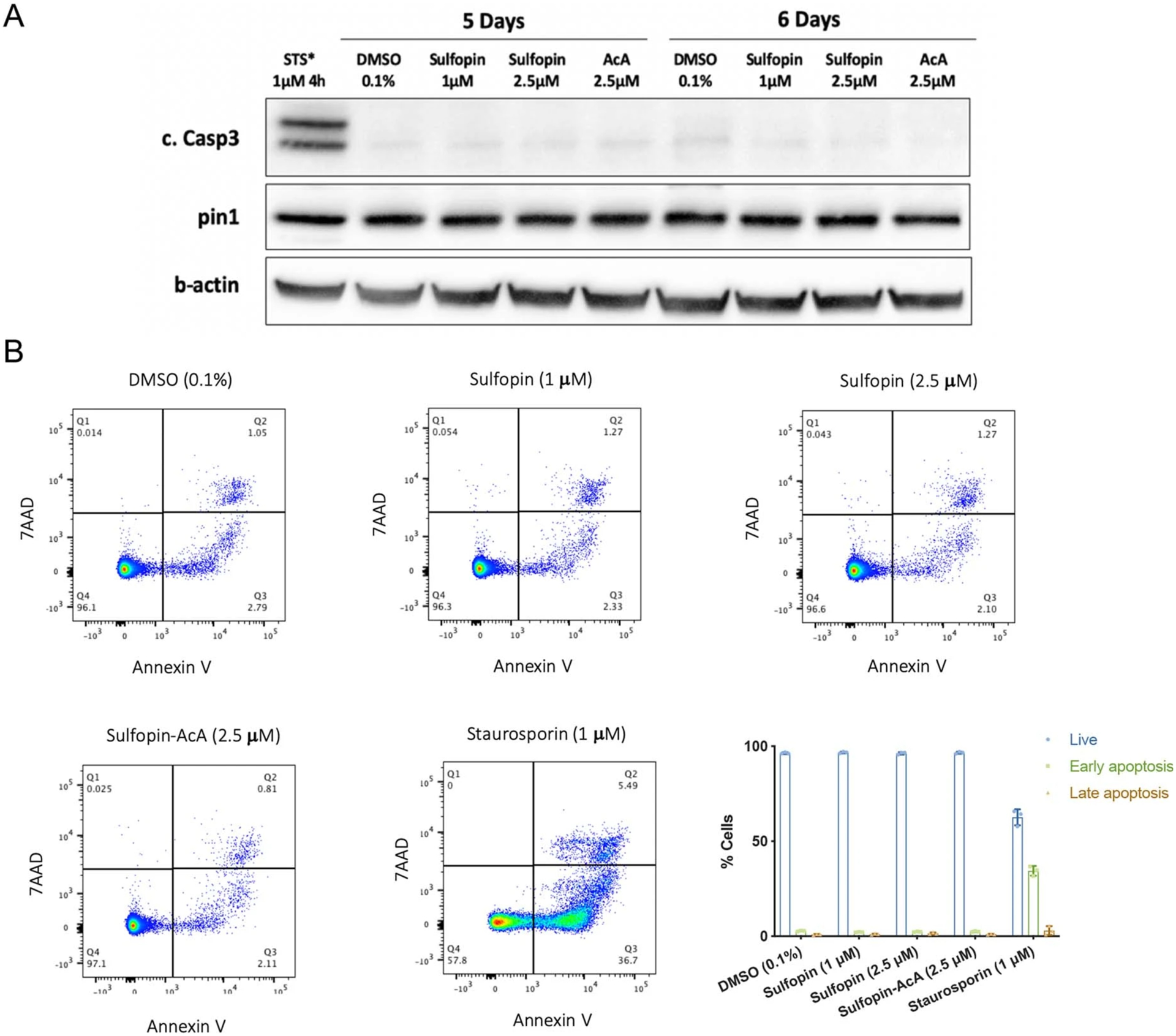
Extended Data Fig. 4 |. Sulfopin treatment does not induce apoptosis in cells.

a, PATU-8988T cells were treated for 5 or 6 days with either DMSO (0.1%), Sulfopin (1 μM, 2.5 μM) or the non-covalent control Sulfopin-AcA (2.5 μM). The cells were lysed and activation of caspase 3 and Pin1 levels were analysed by Western blot. As a positive control for caspase 3 activation the cells were treated with Staurosporin (1 μM, 4h; STS). See Supplementary Fig. 13a for the results of an additional independent experiment. Caspase 3 was not activated and Pin1 levels were not changed by the treatment with Sulfopin. b, PATU-8988T cells were treated in triplicates for 6 days with either DMSO (0.1%), Sulfopin (1 M, 2.5 M) or the non-covalent control Sulfopin-AcA (2.5 M). The cells were then stained with AnnexinV-FITC/ 7AAD and analysed by FACS. Staurosporin treatment (1 M, 4h) was used as a positive control for apoptosis. Representative FACS analysis graphs and a quantification of the results (n=3; data are represented as mean values with standard deviation). See Supplementary Fig. 13b for the results of an additional independent experiment. Live cells were defined as AnnexinV−/7AAD−, early apoptosis AnnexinV+/7AAD− and late apoptosis AnnexinV+/7AAD+.
