
Figure 6. Knock-down of endogenous miR-34a and miR-146b results in up-regulation of tumor suppressors and inhibition of cell proliferation.
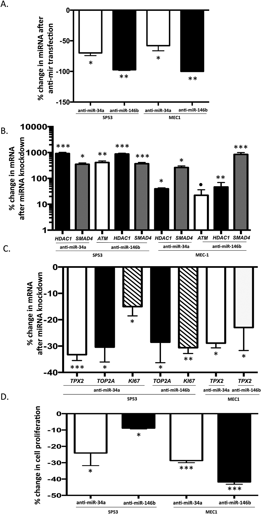
The scrambled control, anti-miR-34a and anti-miR-146b were transfected into SP53 and MEC1 cells, respectively. The expression of miR-34a and miR-146b and putative target mRNA transcripts was measured by quantitative PCR in triplicate. The change in cell proliferation rate was assessed by qPCR for cell proliferation markers and by WST assay. Mean and standard deviation are shown. (a) The expression of miR-34a and miR-146b were significantly decreased compared with the scramble control transfection in both SP53 and MEC1 cells 30 hours after the anti-miR-34a and anti-miR-146b transfection. (b) The expression of the putative target mRNA transcripts of miR-34a and miR-146b was significantly increased in SP53 and MEC1 cells after the anti-miR-34a and anti-miR-146b transfection. (c) The expression of cell proliferation markers (Ki-67, TOP2A, and TPX2) was significantly decreased in SP53 and MEC1 cells after knockdown of miR-34a and miR-146b by anti-miR-34a and anti-miR-146b transfection. (d) Change in cell proliferation is significantly decreased after knockdown of miR-34a and miR-146b as measured by the WST assay 24 h after the anti-miR-34a and anti-miR-146b transfection in SP53 and MEC1 cells. P values were calculated by the Student’s t-test. *, p < 0.05, **, p < 0.01; ***, p < 0.001; ●, p =0.08.