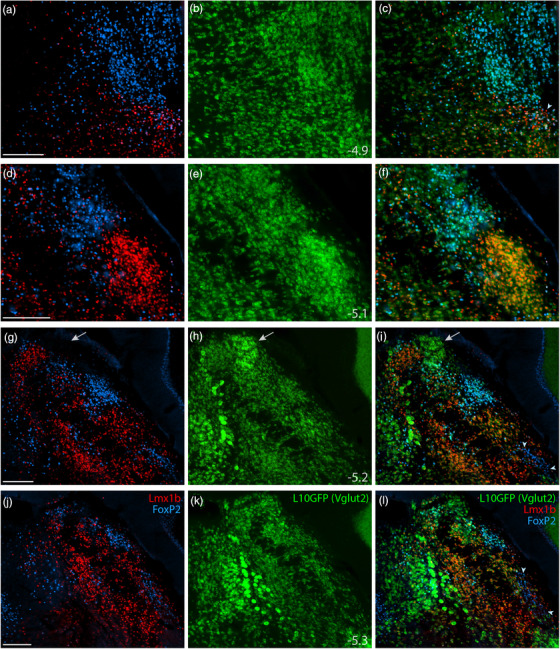
FIGURE 5.

Lmx1b and FoxP2 in a glutamatergic Cre‐reporter mouse. Immunofluorescence labeling identified Lmx1b (red) and FoxP2 (blue) in the PB of mice expressing an L10GFP Cre‐reporter (green) for the glutamatergic marker gene Slc17a6 (vesicular glutamate transporter 2, Vglut2). Arrowhead in (c) indicates a cluster of triple‐labeled neurons (Lmx1b+FoxP2+L10GFP) located rostrally, in the KF region. White arrows in (g–i) highlight a dorsal cluster of L10GFP‐expressing neurons lacking both Lmx1b and FoxP2. Arrowheads in (i) and (l) indicate a ventrolateral cluster of neurons labeled for FoxP2 in the “caudal KF.” Approximate bregma levels are shown at bottom‐right in the center column (in mm). All scale bars are 200 μm and apply to other panels in their respective rows
