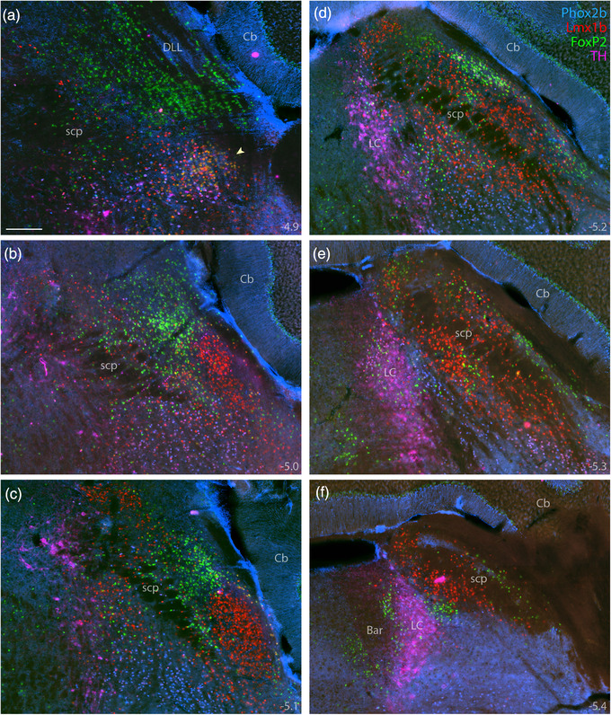
FIGURE 9.

Combined immunofluorescence labeling for Phox2b, Lmx1b, and FoxP2. Immunolabeling for Phox2b (blue, mouse monoclonal antibody) combined with Lmx1b (red) and FoxP2 (green), across six rostral‐to‐caudal sections through the PB region (a–f), distinguished adult PB neurons from underlying non‐PB populations. Additional labeling for TH (magenta) distinguished Phox2b neurons in the LC from those in the supratrigeminal population. Approximate bregma levels are shown at the bottom‐right of each panel. Arrowhead in (a) highlights the KF. Scale bar in (a) is 200 μm and applies to all panels
