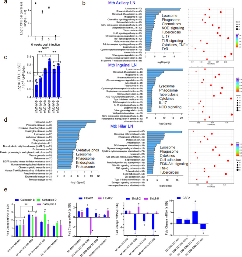
Fig. 7. Inreg clusters expressed by Mtb-infected human M1- and M2-MФs are found in the lymph nodes and macrophage transcriptome of Mtb-infected neonatal rhesus macaques.
a Six-week-old rhesus macaques were aerosol-infected with Mtb Erdman strain (25 CFU per animal; n = 4) followed by sacrifice at 6 weeks and CFU counts of lungs. b lymph nodes (n = 2) collected at necropsy from macaques that had comparable Mtb counts in lungs (panel a) were analyzed using RNAseq. Kyoto Encyclopedia of Genes and Genomes profiles of one NHP illustrate the differential gene expression for Inreg clusters; Clusterprofiler pathway analysis of Inregs is illustrated in Supplementary Fig. 12. c M1-, M2-, and M0-MΦs were prepared from the bone marrow of naïve macaques (prefix n) and infected with Mtb followed by CFU counts on day 4 (**p < 0.01 one-way ANOVA with Tukey’s post-hoc test; 2 experiments shown). d Kyoto Encyclopedia of Genes and Genomes profiles of Mtb-infected M1-MΦs vs. Mtb-infected M2-MΦs show enrichment of genes regulating antigen processing 18 h post-infection. e Naïve or Mtb-infected M1-, and M2-MΦs were subjected to QPCR at 18 h post-infection using primers for mRNA of indicated genes which were differentially expressed in their human counterparts (*p < 0.01, t test). All ex vivo Mtb CFU experiments used MOI of 1.

