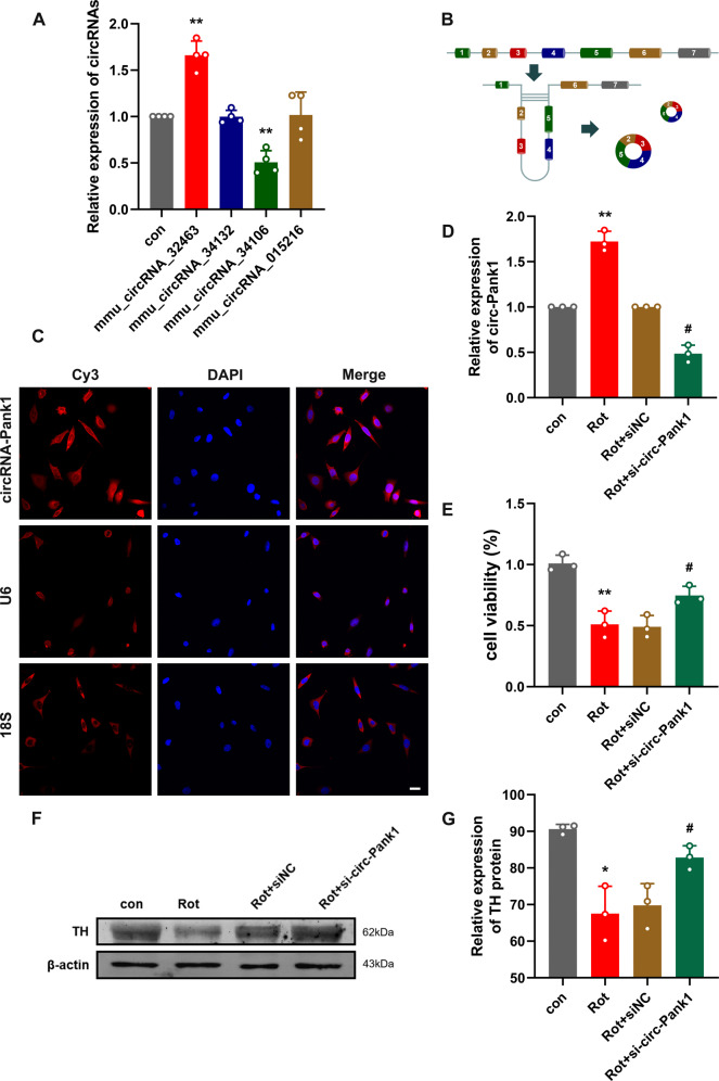
Fig. 2. The expression of circ-Pank1 significantly increased in PD model mice treated with rotenone and downregulation of circ-Pank1 in the MN9D cell model alleviates the damage to dopaminergic neurons.
A qRT–PCR was used to detect the expression of mmu_circ_32463, mmu_circ_34132, mmu_circ_34106, and mmu_circ_015216 in the SN of PD model mice treated with rotenone (n = 4). B Schematic diagram of ring formation of circ-Pank1. C Sublocalization of circ-Pank1 in MN9D cell. Taking U6 in the nucleus and 18S in the cytoplasm as the controls, most circ-Pank1 in MN9D cells was located in the cytoplasm; scale bars, 50 μm. D–G After si-circ-Pank1 and the corresponding control were transferred into the MN9D cell model treated with rotenone (n = 3). D qRT–PCR was used to detect the expression of circ-Pank1. E CCK-8 was used to detect the cell viability. F, G Western blot analysis of the TH protein levels. Data are presented as mean ± SD. *P < 0.05, **P < 0.01.

