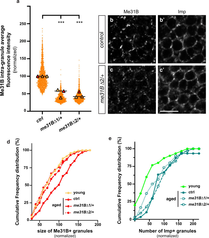
Fig. 5. Reducing Me31B levels decreases Me31B condensation.
a Intra-granule concentration of Me31B-GFP in granules of 37–38 day-old me31B Δ1/ + and me31BΔ2/ + MB γ neurons. Three replicates were performed and the mean value of each replicate is indicated as a symbol (triangle). Values were normalized to the control condition. The distribution of individual granule values is shown for one replicate only. ***P < 0.001 (one-way ANOVA with Dunn’s multiple comparison tests). b, c Cell bodies of control (b, b’) and me31BΔ2/ + (c, c’) MB γ neurons from 37–38 day-old brains stained with anti-Me31B (b, c) and anti-Imp (b’,c’). Scale bar: 5 μm. d Sizes of Me31B-containing granules in MB γ neurons of 1–2 day- (light orange; young) and 37–38 day- (red; aged) old brains. Values were normalized to aged controls. Cumulative frequency distributions are plotted for one replicate, but three replicates were performed in total (see Supplementary Fig. 4d). ***P < 0.001 (one-way ANOVA test). e Numbers of Imp + granules (per surface area) in MB γ neurons of 1–2 day- (light green; young) and 37–38 day- (green; aged) old brains. Values were normalized to aged controls. Cumulative frequency distributions are plotted for one replicate, but three replicates were performed in total (see Supplementary Fig. 4f). ***P < 0.001 (one-way ANOVA test). Source data are provided as a Source Data file.

