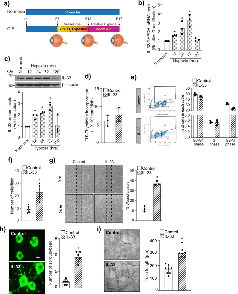
Fig. 1. IL-33 induce sprouting angiogenesis in retinal endothelial cells.
a Schematic diagram representing the murine OIR model. C57BL/6 mice pups with dams were exposed to 75% oxygen from P7 to P12 and returned to room at P12. At P17, eyes were enucleated, retinas isolated. b Total cellular RNA was isolated from retina and quantified for IL-33 and GAPDH mRNA levels by QRT-PCR. c Retinal tissue extracts were prepared and analyzed for IL-33 by Western blotting and normalized to β-tubulin. n = 6 mice per group. Quiesced HRMVECs were treated with and without IL-33 (20 ng/mL) and cell proliferation was measured by thymidine incorporation (d), and BrdU cell proliferation assay (e). f, g Everything is same as in d, except that cell migration was measured using Boyden chamber method (f) and wound healing assay (g). h HRMVECs cells were labeled, coated onto cytodex beads, embedded in a 3D-fibrin gel and sprouting was observed after 3 days under Zeiss LSM800 microscope. i Everything is same as in d, except that tube formation was assessed using growth factor reduced Matrigel. The bar graphs show the quantitative analysis of three independent experiments, expressed as Mean ± SD. *P < 0.05 vs control or normoxia. Scale bar represents 50 μm in h.

