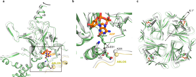
Fig. 3. Conformational changes of Kir6.2 CTD during KATP channel opening.
a Conformational changes of the nucleotide-binding site between the closed (gray) and the pre-open (green) states of H175Kcryo-EM. The ADP bound in the closed state is shown as sticks in orange. b Close-up view of the nucleotide-binding site boxed in (a). c Bottom view of the Kir6.2 CTD. The rotation angle between CTDs was measured using Cα positions of L356 of Kir6.2 as marker atoms (shown as spheres).

