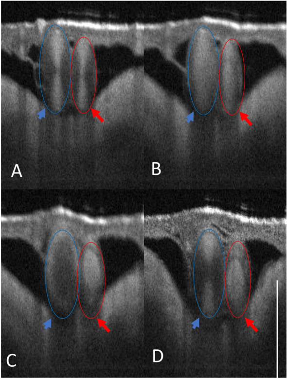
Figure 3.

OCT B-scan image of CAM showing an arteriole (red)/venule (blue) pair irradiated at the same fluence rate. (A) Initial OCT B-scan before laser irradiation. (B) Coagulum formation observed in both arterioles and venules. (C) In the arteriole, the coagulum holds, resulting in hemostasis, while in the venule, the coagulum breaks free near the wall. (D) Blood flow is restored in the venule where the coagulum breaks from the vessel wall (Panel C). Please see supplementary Fig. S10 for enface images collected from this experiment. Scale bar 500 µm. These arteriole/venule pairs were chosen on the CAM model appropriately sized at Day 7 with direct connection to major CAM vessels.
