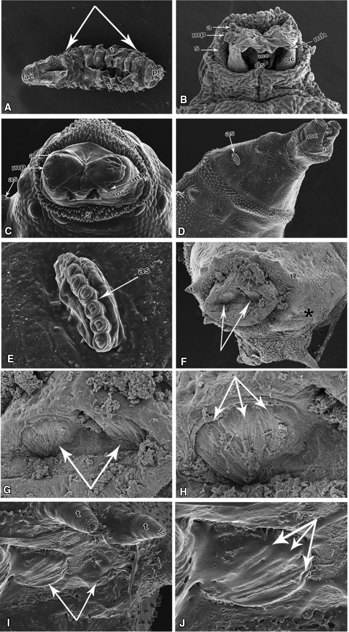
Figure 4.
Scanning electron micrographs of a third instar larva of Chrysomya albiceps from the AlP group. (A) Larval body composed of groups of tubercles (t) located at the anterior and posterior ends of each segment, anterior (ae), and posterior ends (pe). Notes: Shrinkage in larval length, arrows abdominal segments. (B) Ventral view of the cephalic region with antennae (a), maxillary palp (mp), spines (s), dental sclerite (ds), short mouth hooks (mh), oral cristae (oc). (C) Dorsal view of the cephalic region with antennae (a), maxillary palp (mp), spines (s), dental sclerite (ds), short mouth hooks (mh), oral cristae (oc), and deformed anterior spiracle (as). (D) Details of antennae (a) and maxillary palp with five papillae and deformed anterior spiracle (as), spines (s), oral cristae (oc), and dental sclerite (ds). (E) Details of anterior spiracles (as) in a row. (F) Anal segment with posterior spiracles (arrows), tubercles (t). (G) Magnified part of the micrograph. (F) Details of posterior spiracles. (H) Details of the anal segment with deformed three spiracular openings (arrows). (I) Hypogenesis of posterior spiracles (arrows). (J) Completely deformed three spiracular openings (arrows).

