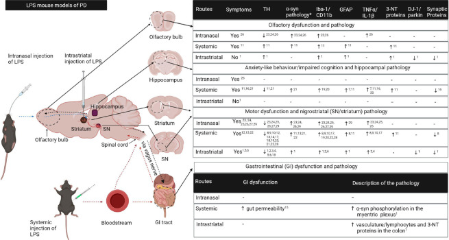
Figure 1.
A summary of the pathological changes in LPS models of PD.
Intranasal, systemic and intrastriatal administration of LPS induce pathological changes in the olfactory bulb, hippocampus, striatum, substantia nigra (SN) and the gastrointestinal tract which are associated with functional changes similar to clinical PD. Blue dotted arrows indicate propagation of LPS associated signals from the olfactory bulb to the nigrostriatal pathway; red dotted arrows – from periphery to the nigrostriatal pathway; and the blue dotted arrows – from the striatum to the olfactory bulb and gastrointestinal (GI) system. The parameters in the first row of the table apply to the olfactory bulb, hippocampus and striatum/SN but each parameter must be read vertically; α-syn pathology* refers to the increased α-synuclein protein expression, phosphorylation and/or aggregation; “-” means “not examined”. 3-NT: 3-nitrotyrosine.; α-syn: α-synuclein; DJ-1: deglycase DJ-1; GFAP: glial fibrillary acidic protein; Iba-1: Ionised calcium-binding adaptor molecule 1; IL-1β: interleukin-1β; PD: Parkinson’s disease; SN: substantia nigra; TH: tyrosine hydroxylase; TNF-α: Tumour necrosis factor-α. References: 1 (Deng et al., 2021b); 2 (Garcia et al., 2018); 3 (Gao et al., 2008); 4 (Gómez-Gálvez et al., 2016); 5 (Hunter et al., 2009); 6 (Zhang et al., 2012); 7 (Badshah et al., 2016); 8 (Beier et al., 2017); 9 (Bodea et al., 2014); 10 (Deneyer et al., 2019); 11 (Deng et al., 2021a); 12 (Jiang et al., 2017); 13 (Liu et al., 2008); 14 ( Milde et al., 2021); 15 (Kelly et al., 2014); 16 (Khan et al., 2019); 17 (Qin et al., 2013); 18 (Qin et al., 2004; 19 (Qin et al., 2007); 20 (Song et al., 2019); 21 (Zhang et al., 2019); 22 (Zheng et al., 2013); 23 (He et al., 2013); 24 (He et al., 2016); 25 (Li et al., 2015); 26 (Niu et al., 2020); 27 (Zhao et al., 2018); 28 (Liao et al., 2021); 29 (Song et al., 2020). The figure was created with BioRender.com.

