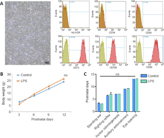
Figure 1.

Experimental flowchart and growth and development of newborn pups with maternal immune activation-associated neonatal brain injury.
(A) Cell morphology and surface markers of passage 5 cells. Umbilical cord mesenchymal stem cells were spindle-shaped. CD73, CD105, and CD90 staining was positive, with positive expression rates of 99.94%, 98.62%, and 99.86%, respectively. HLA-DR, CD34, and CD45 showed expression rates of 0.13%, 0.36%, and 0.18%, respectively. Scale bar: 250 μm. (B) Neonatal rat weight was measured every 3 days. (C) Assessment of developmental indicators of neonatal rats. The pups were tested for ear standing, righting reflex, incisor odontogenesis, auditory astonishment, and eye opening. Data are expressed as mean ± SEM (control: n = 13, LPS + PBS: n = 11, LPS + UC-MSC: n = 9). ns: Not significant, vs. control group (one-way analysis of variance followed by the Student-Newman-Keuls method). LPS: Lipopolysaccharides; P: postnatal day; PBS: phosphate-buffered saline; UC-MSCs: umbilical cord-derived mesenchymal stem cells.
