
Figure 2.
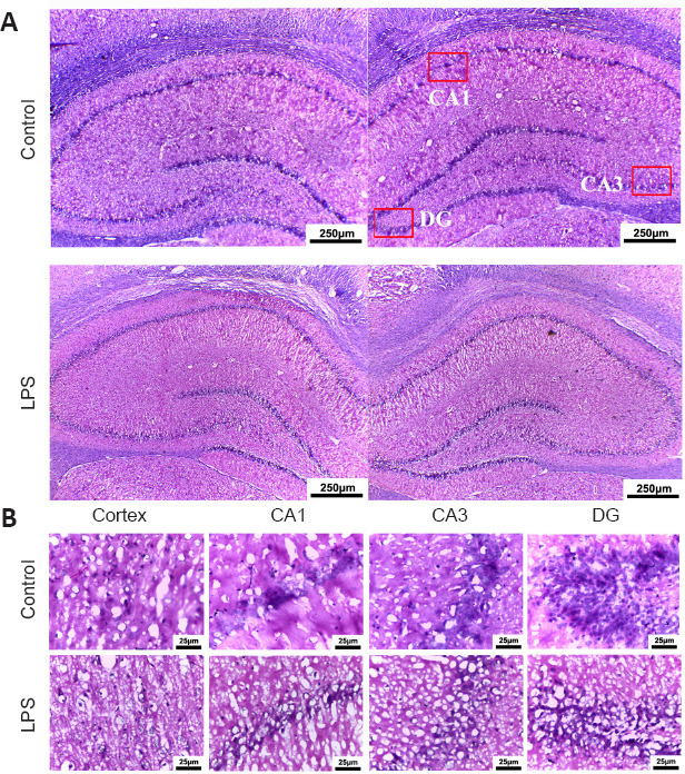
Pathological changes in the brain of neonatal rats with maternal immune activation-associated neonatal brain injury (hematoxylin and eosin staining).
(A) Pathological changes in neonatal rats’ hippocampus (original magnification 40×, scale bars: 250 μm). The cortical cells in the LPS group had obvious nucleoli, the number of cells was significantly reduced, and the arrangement was loose, showing a “vacuum” appearance. The hippocampal structure in the control group was more intact and cells were lined up more neatly. (B) Pathological changes in the cortex and hippocampal CA1, CA3, and DG (original magnification 200×, scale bars: 25 μm). Compared with the control group, the number of neurons in the brain cortex and hippocampus of young rats in the LPS group was significantly reduced. The neuronal nuclei were deeply stained and the nucleoli disappeared. Moreover, the neuronal structure was loose and lacked hierarchy. DG: Dentate gyrus; LPS: lipopolysaccharides; PBS: phosphate-buffered saline; UC-MSCs: umbilical cord-derived mesenchymal stem cells.