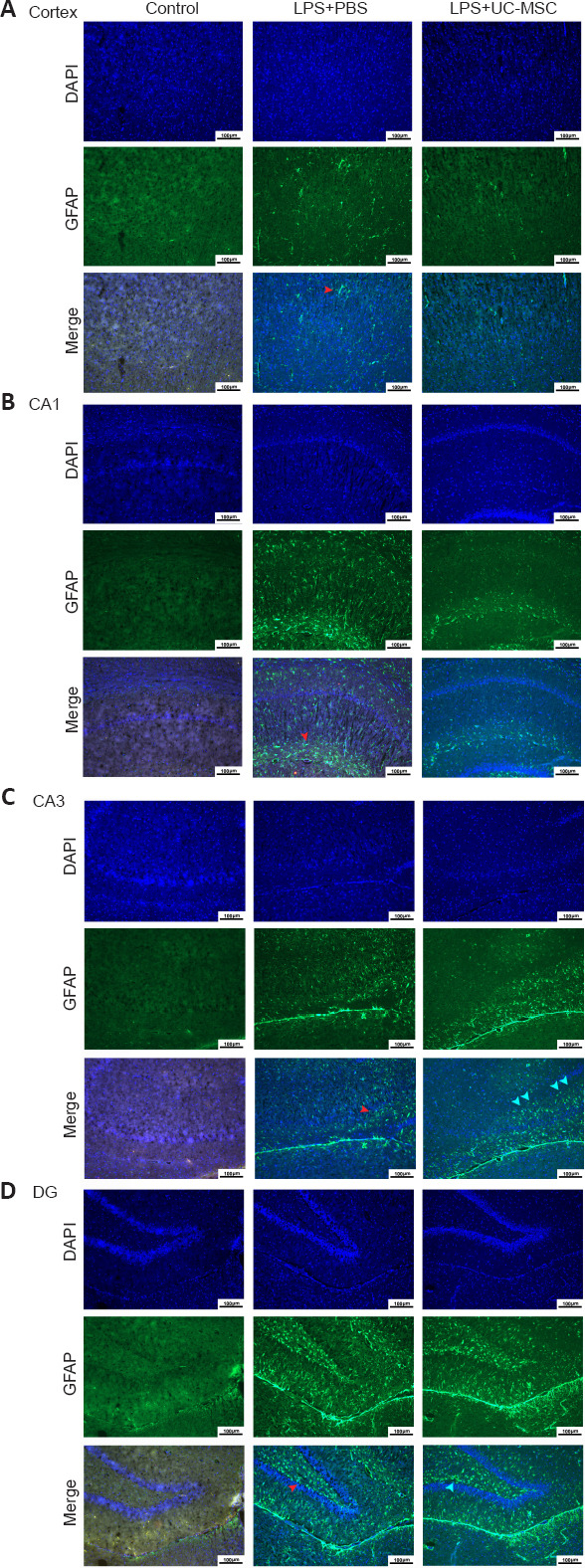
Figure 5.

Effects of UC-MSCs on the distribution of GFAP-positive DAAs in the cerebral cortex (A) and hippocampus CA1 (B), CA3 (C), and DG (D) of neonatal rats with maternal immune activation-associated neonatal brain injury (immunofluorescence).
In the LPS + PBS group, the GFAP (green, stained by FITC)-positive activated astrocytes (red arrows) located in the cortex were arranged in a centripetal pattern and were more evenly distributed in the hippocampus CA1, CA3, and DG. In the LPS + UC-MSC group, GFAP-positive activated astrocytes were densely distributed along the hippocampal projection pathway (green arrow). Original magnification 100×, scale bars: 100 μm. DAPI: 4′,6-Diamidino-2-phenylindole; DG: dentate gyrus; GFAP: glial fibrillary acidic protein; LPS: lipopolysaccharides; PBS: phosphate-buffered saline; UC-MSCs: umbilical cord-derived mesenchymal stem cells.
