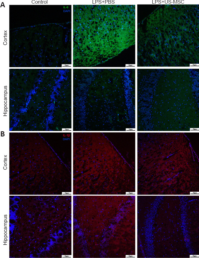
Figure 6.

Effects of UC-MSCs on the immunopositivity of inflammatory cytokines IL-6 (A) and IL-1β (B) in the cerebral cortex and the hippocampus of neonatal rats with maternal immune activation-associated neonatal brain injury (immunofluorescence).
IL-6 (green, stained with FITC) was highly expressed in the cortex of the LPS + PBS group. In the LPS + UC-MSC group, the immunopositivity of IL-6 in the cortex was lower than that in the LPS + PBS group. There was no significant difference between these groups in the immunopositivity of IL-6 in the hippocampus. Compared with the control group, IL-1β expression (red, stained with Alexa Fluor 647) was higher in the cortex and hippocampus of the LPS + PBS group. In the PBS + UC-MSC group, the immunopositivity of IL-1β in the cortex was similar to that in the LPS + PBS, whereas in the hippocampus it was lower. Original magnification 100×, scale bars: 75 μm. DAPI: 4′,6-Diamidino-2-phenylindole; DG: dentate gyrus; IL-1β: interleukin-1β; IL-6: interleukin-6; LPS: Lipopolysaccharides; PBS: phosphate-buffered saline; UC-MSCs: umbilical cord-derived mesenchymal stem cells.
