Fig. 1.

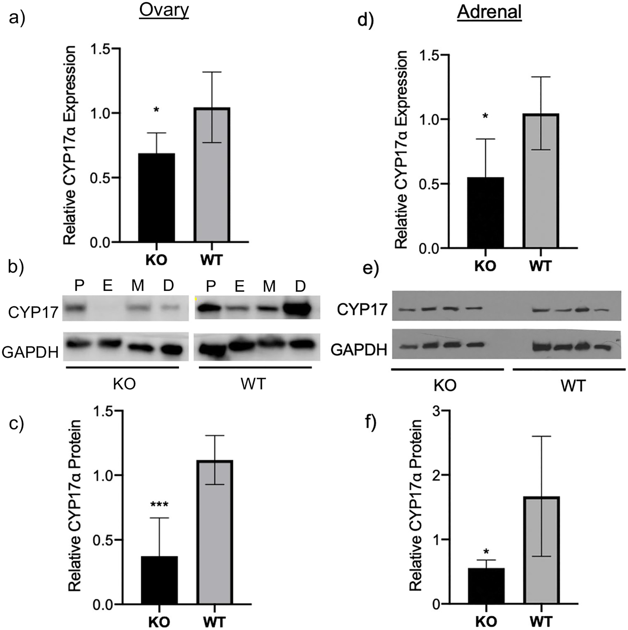
Decreased CYP17α levels in ovaries of H19KO female mice. To evaluate CYP17α expression, RNA extraction, cDNA synthesis, and RT-qPCR were performed on ovarian tissue from 8-week-old H19KO and WT female mice (n = 5 per group). a Mean relative Cyp17α expression levels by real-time PCR are shown. Cyp17α expression was decreased in ovaries from H19KO female mice compared to that from WT. Beta-actin was used as a control. b Western blot analysis was performed to measure CYP17α protein levels, using GAPDH as control. c Quantification of Western blot showing decreased CYP17α protein in ovary compared to that in WT. All data were analyzed using Student’s t-test. *p = 0.05; ***p < 0.001. Error bars represent one SEM. P, proestrus; E, estrus; M, metestrus; D, diestrus
