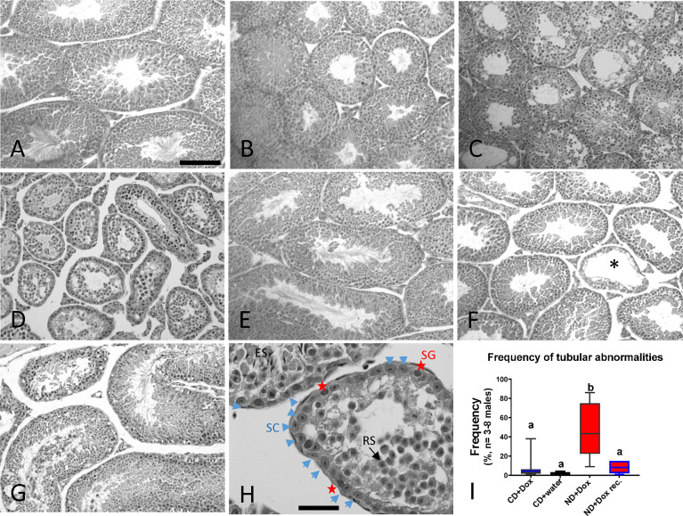
Figure 3.
Impact of testicular NAD+ levels and male age on the seminiferous epithelium. Hematoxylin/eosin-stained testicular sections of testes from young adult male fed (A) CD+Dox control diet, (B) ND+Dox diet for 8 weeks, (C) ND+Dox for 14 weeks; damaged tubules (D) ND+Dox for 24 weeks; normal tubules +/- absent (E) ND for 24 weeks followed by 9 weeks of recovery on niacin-containing CD diet; tubules mostly restored. (F) Control testis at 20 months of age. Asterisk marks a tubule with abnormal seminiferous epithelium. (G) Control testis of 31 month-old mouse. (H) After 24 weeks on ND diet, seminiferous tubules are lined mostly by Sertoli cells (SC, blue arrow heads) interspersed with spermatogonia (SG, red stars), as identified by histological morphology of the cells. Tubular lumen contain mostly cells resembling round spermatids (RS, black arrow) and occasionally elongated spermatids (ES)). (I) NAD+-deficient testis contain significantly more tubules with abnormal composition of the seminiferous epithelium in testis of mice that were on indicated diets for 16 weeks (CD+Dox, CD+water, ND+Dox) or ND+Dox that were subsequently recovered on niacin containing diet for 9 weeks. One hundred tubules were evaluated per testis section, ANOVA with Tukey’s multiple comparison, b is significantly different from a, p=0.0003 to 0.001. Scale bar: 250 mm in a.-f., 40 mm in (g) & h.

