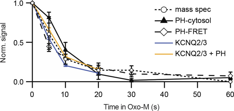
Figure 4.
Depletion of PtdIns(4,5)P2 in response to a muscarinic agonist (Oxo-M) in cell lines. The diamonds show loss of FRET when FRET pairs PHPLCδ1-CFP and PHPLCδ1-YFP translocate from being bound near each other at the plasma membrane to being free in the cytoplasm, and the triangles show appearance of PHPLCδ1-YFP in the cytoplasm from confocal images (transfected tsA201 cells). Circles are PtdInsP2 from mass spectrometry (mass spec, in CHO-M cells stably expressing M1 receptors), and colored lines are amplitudes of KCNQ2/3 current (tsA201 cells; Jensen et al., 2009; from Traynor-Kaplan et al., 2017).

