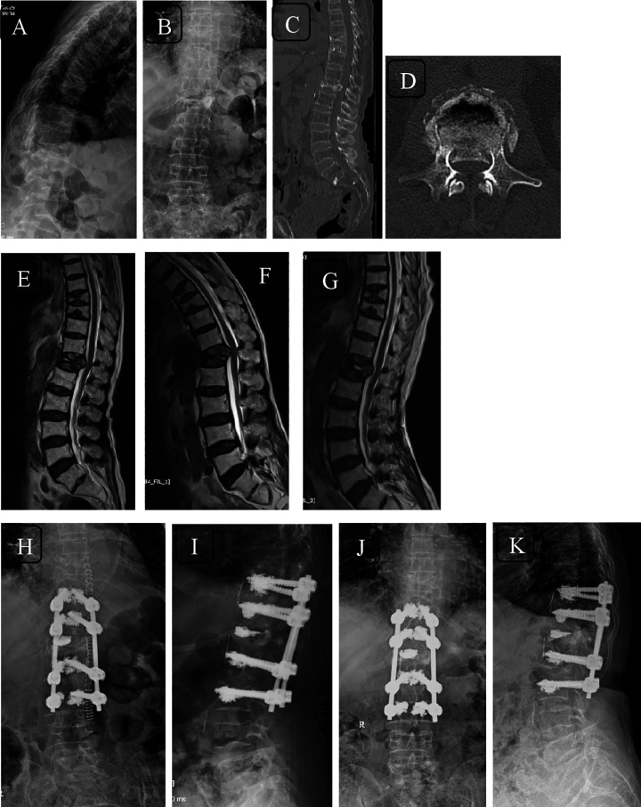
Figure 5.
Radiographs (A, B) and computed tomography (CT) scans (sagittal—C; axial—D) of a 61-year-old woman with fracture T12 with incomplete neurology with canal compromise evident on CT and neutral magnetic resonance imaging (MRI) (E). The dynamic nature of compression on spinal cord can be appreciated on flexion MRI (F) and canal clearance on extension MRI (G). Immediate postoperative images (H, I) and 2-year follow-up images (J, K) showing cement augmented long segment fixation.

