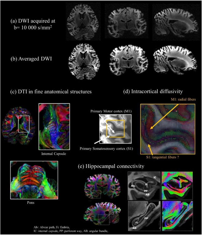
Fig. 15. Whole-brain ex-vivo diffusion MRI at 550 micrometer isotropic resolution using b-values up to 10000s/mm2.
Axial, coronal, and sagittal views of a given diffusion direction are shown in (a), and the corresponding averaged DWI are displayed in (b). High-quality submillimeter diffusion MRI allows mapping diffusivity with unprecedented quality in fine anatomical structures often inaccessible in in-vivo settings, as seen in the internal capsule and transverse fibers in the pons (c), and anisotropic diffusivity in the primary and somatosensory cortex (d). Resolving fiber architecture of the hippocampus is achievable using this high-quality, high-spatial-resolution dataset, as can be seen in (e). Adapted from (Ramos-Llorden et al., 2021).

