
Fig. 4 –
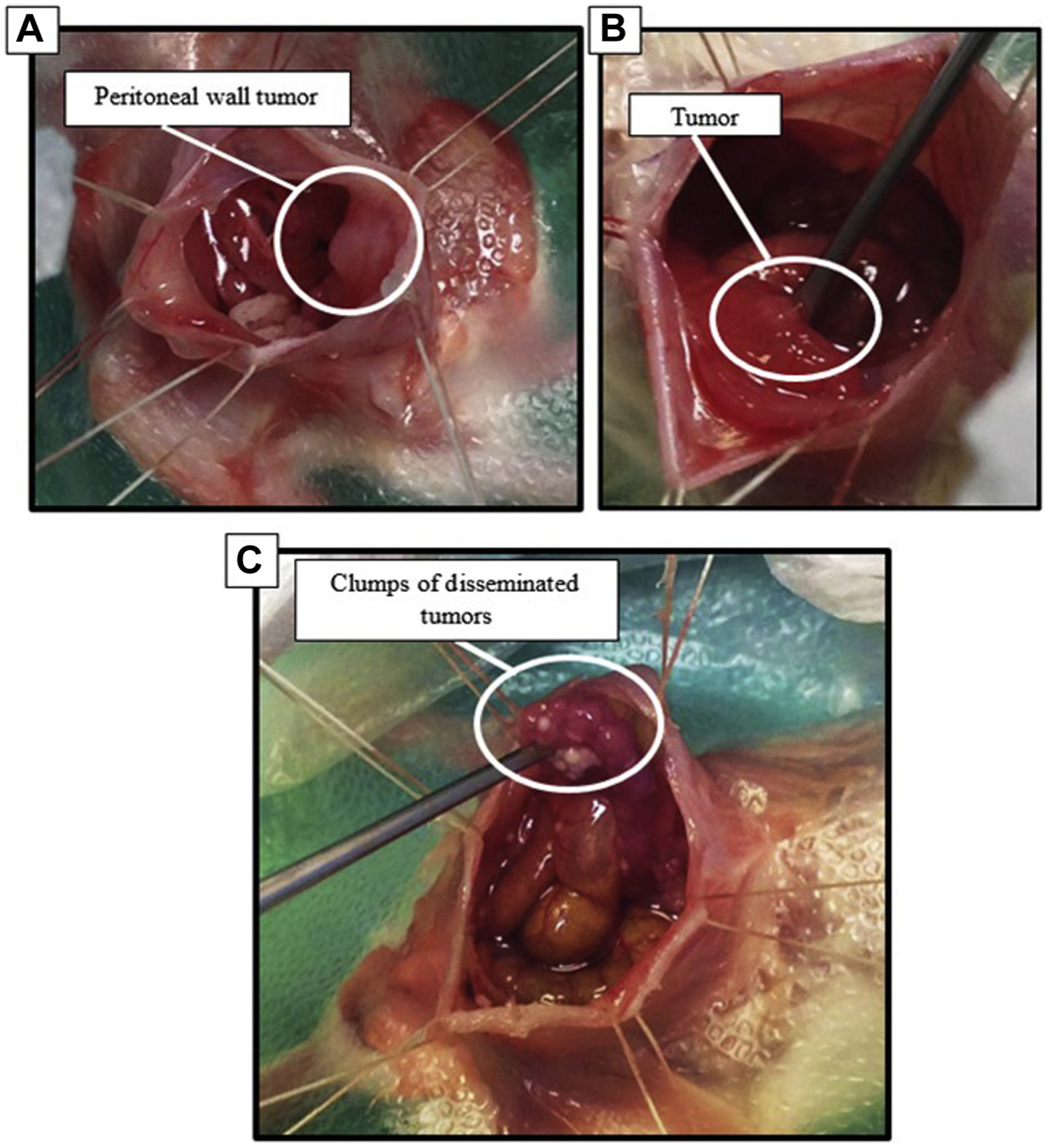
Tumors that developed in IP injection dissemination model. (A) Tumors adhered to internal peritoneal wall (B) tumors integrated onto surface of the large intestine. (C) Clumps of tumors with poor vasculature; these tumors were easily disturbed and produced mucus.