Figure 2.
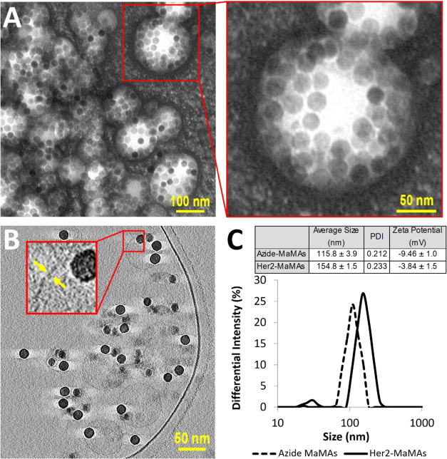
(A) TEM images of MaMAs obtained using negative staining with 2% uranyl acetate. (B) Cross-sectional cryo-EM images of MaMAs. Individual IONPs (25 nm in diameter) can be clearly seen within spherical structures. Note the absence of a visible lipid bilayer, indicating that these structures are not liposomes. (C) Size (DLS, intensity) and ζ-potentials of MaMAs before and after conjugation with trastuzumab.

