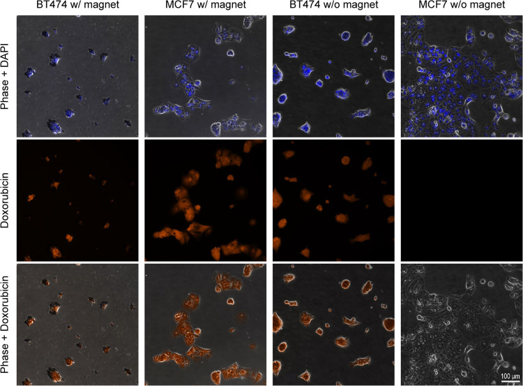
Figure 6.
HER2+ BT474 and HER2– MCF7 cells after incubation with aHER2-DOX-MaMAs at 37 °C with or without a permanent magnet: (top row) combined phase and DAPI images; (middle row) fluorescence images of doxorubicin; and (bottom row) combined phase and doxorubicin images. The images were acquired using a Zeiss Axio Observer.Z1m microscope equipped with a Hamamatsu ORCA-ER camera (Bridgewater, NJ) under a 40× objective lens. Fluorescence images were obtained with BP 550/25 nm excitation and BP 605/70 nm emission filters for doxorubicin detection and G 365 nm excitation, BP 445/50 emission filters for DAPI. Scale bar is 100 μm for all images.

