
FIGURE 1.
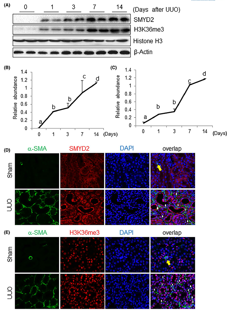
Time dependent SMYD2 expression and H3K36 trimethylation in the kidney after obstructed kidneys. A, Kidneys were collected at different time points as indicated from sham-operated or obstructed kidneys of mice and the prepared tissue lysates were subjected to immunoblot analysis with antibodies against SMYD2, H3K36me3 or β-actin (A). The levels of SMYD2, H3K36me3, and β-actin were quantified by densitometry; SMYD2 and H3K36me3 levels were normalized to β-actin and Histone H3, respectively (B, C). Values are the means ± SDs (n = 6). Bars with different letters (a-d) are significantly different from one another (P < .05). D, E, Photomicrographs illustrate co-staining of α-SMA and SMYD2 (D) or H3K36me3 (E) in the tissue section of the obstructed kidney (magnification ×600). DAPI: 4’,6-diamidino-2-phenylindole. The yellow arrow indicates small blood vessels, and white arrows indicate SMYD2 or H3K36me3-positive myofibroblasts