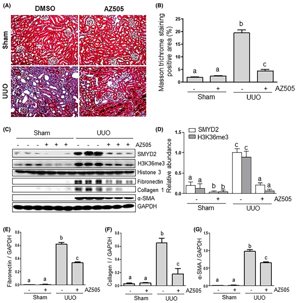
FIGURE 2.

AZ505 attenuates development of renal fibrosis and deposition of ECM in obstructed kidneys. A, Photomicrographs illustrating Masson trichrome staining of kidney tissue (magnification ×200). B, The Masson trichrome-positive tubulointerstitial area (blue in A) relative to the whole area from ten random cortical fields was analyzed. Data are represented as the means ± SDs (n = 6). C, Kidney tissue lysates were subjected to immunoblot analysis with antibodies against SMYD2, H3K36me3, Histone H3, α-SMA, collagen 1, fibronectin, or GAPDH (C). Expression levels of SMYD2, H3K36me3, fibronectin, collagen 1, α-SMA, or GAPDH were quantified by densitometry, and the levels of SMYD2 (D), fibronectin (E), collagen I (F), and α-SMA (G) were normalized with GAPDH. H3K36me3 levels were normalized with Histone H3 (D). Values are the means ± SDs (n = 6). Means with different letters (a-c) are significantly different from one another (P < .05)
