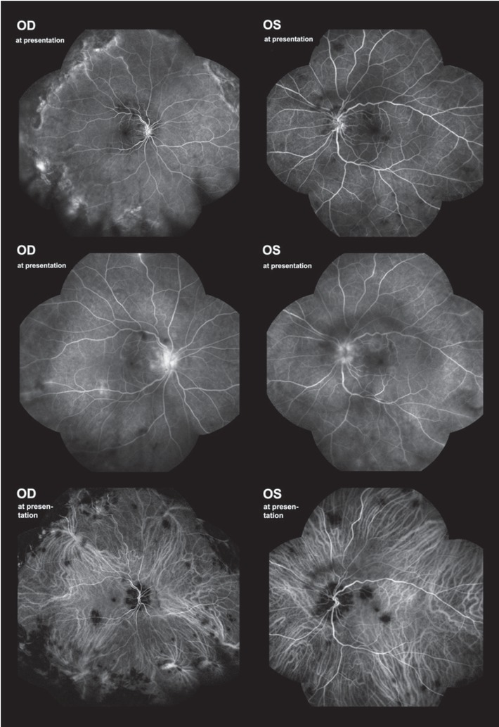
Figure 7.

Case II. Fluorescein angiography at presentation, early and late frames. Note surface capillary net dilatation and late staining of the papilla and hypofluorescent areas on the posterior pole and periphery. Indocyanine angiography at presentation showing hypofluorescent lesions.
OD = oculus dexter; OS(L) = oculus sinister
