. 2022 May 20;11:e72534. doi: 10.7554/eLife.72534

Figure 2. Determination of viral vectors for long-range anterograde tracing in macaques.

(A) GFP-labeled neurons were found in the premotor cortex ~5 days after injection of VSV-△G encoding Tau-GFP. (B) A magnified view illustrating the morphology of GFP-labeled neurons in the area outlined with a white box in (A). (C) Lentivirus construct was injected into the macaque thalamus and examined for transgene expression after ~9 months. (D) High power views of the dotted rectangle in panel C. (E) GFP-labeled neurons and axons were observed in the premotor cortex ~45 days after injection of AAV2/9 encoding Tau-GFP. Two dashed line boxes enclose the regions of interest: frontal white matter and ALIC, whose GFP signal are magnified in (F) and (G), respectively.

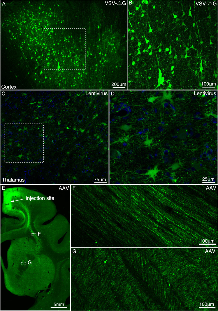
Figure 2.

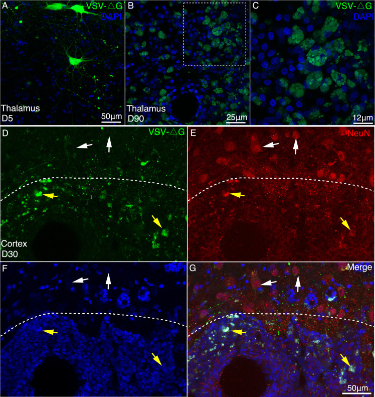
Figure 2—figure supplement 1. Long-term expression of VSV-△G induced neurotoxicity in macaque brain.

Figure 2—figure supplement 1.

(A–C) Compared with the short-term expression (A, 5 days), severe morphological abnormalities such as neurites loss and membrane blebbing (C), were induced by VSV (△G) after long-term expression (90 days). (D–G) 30 days after injection of VSV, immunofluorescent staining was performed with NeuN antibody in the injection site (below the dotted line) and noninjection site (above the dotted line). The noninfected neurons were found to be GFP negative and neuronal-specific nuclear protein (NeuN) positive by immunofluorescent examination. In the injection site, VSV-infected neurons with GFP and NeuN positive exhibited apparent morphological abnormalities.
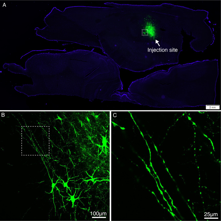
Figure 2—figure supplement 2. Expression of GFP using VSV-△G injected into the MD thalamus of macaque brain.

Figure 2—figure supplement 2.

(A) GFP-labeled neurons were found in the MD thalamus ~5 days after injection of VSV-△G encoding Tau-GFP. (B) A magnified view illustrating the morphology of GFP-labeled neurons in the area outlined with a white box in (A). (C) Higher magnification view of GFP-positive axons.
Figure 2—figure supplement 3. Expression of GFP using lentivirus injected into the MD thalamus of macaque brain.

Figure 2—figure supplement 3.

(A) Lentivirus construct was injected into the macaque thalamus and examined for transgene expression after ~9 months. (B) Magnified view of panel A. (C) High power view of the dotted rectangle in panel B. Note the presence of GFP-positive cells.
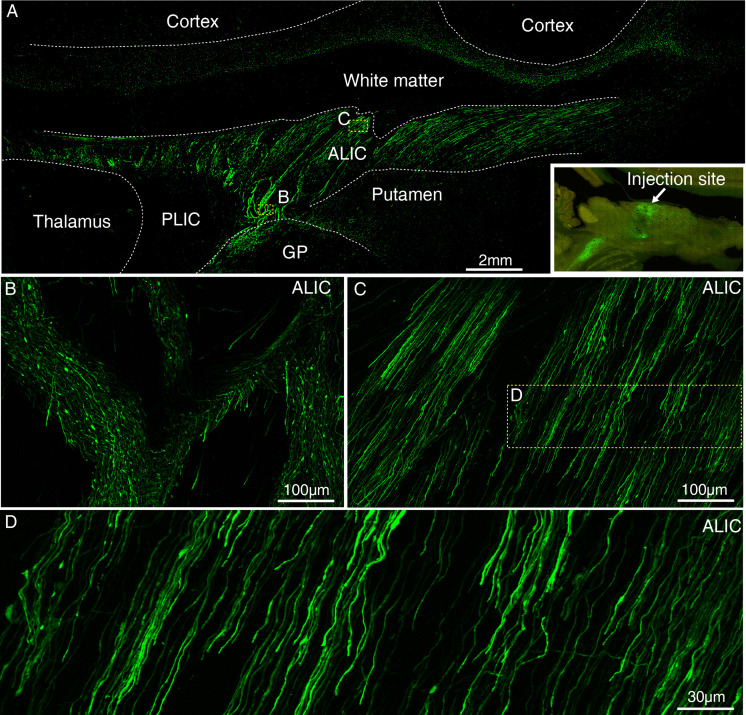
Figure 2—figure supplement 4. Expression of GFP using AAV2/9 injected into the MD thalamus of macaque brain.

Figure 2—figure supplement 4.

(A) GFP-labeled axons were observed in the subcortical regions ~ 45 days after injection of AAV2/9 encoding Tau-GFP in MD thalamus. The inset shows the injection site in MD thalamus. Two dashed line boxes enclose the region of ALIC, whose GFP signal are magnified in (B) and (C). (D) Higher magnification view of GFP-positive axons.
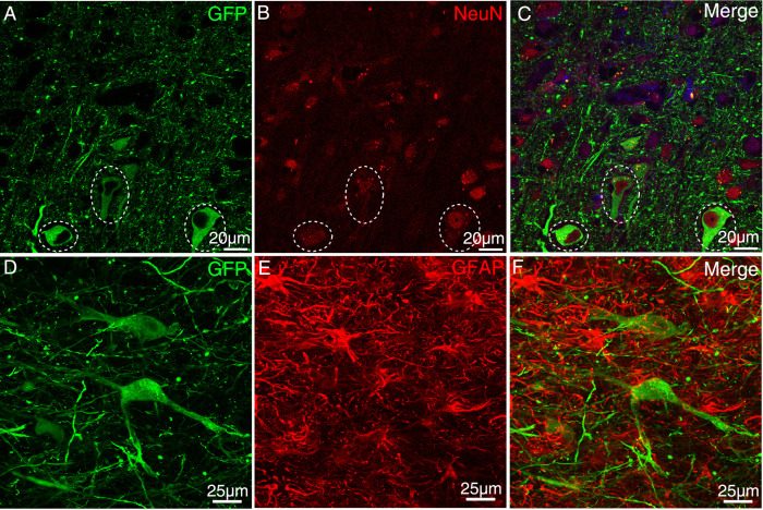
Figure 2—figure supplement 5. AAV-infected cells were stained for NeuN and GFAP antibodies.

Figure 2—figure supplement 5.

After injection of AAV2/9, immunofluorescent staining was performed with NeuN (A–C) and GFAP (D–F) antibodies in the injection site. We found GFP-positive cells positive for the neuronal specific marker NeuN but negative for astrocyte specific marker GFAP.
Figure 2—figure supplement 6. Comparison of two AAV constructs.

Figure 2—figure supplement 6.

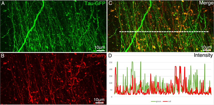
AAV2/9 encoding Tau-GFP and AAV2/9 encoding mCherry were co-injected in the premotor cortex. Figures (A and B) show the axon fibers labeled by Tau-GFP and mCherry, respectively. (C) Colocalization of mCherry and GFP in the axonal fibers. (D) The intensity profiles (measured using ImageJ on 8-bit TIF images) along the dashed line (in C) in red and green channels. After normalization, a direct comparison indicates that the intensity of Tau-GFP was stronger than that of mCherry.
Figure 2—figure supplement 7. Long-range axonal tracing outcomes using AAV2/9, lentivirus,` and VSV-△G.

Figure 2—figure supplement 7.

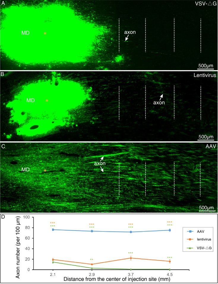
(A) VSV-△G rarely labeled long-range axonal fibers issued from MD thalamus. (B) Only sparse axon fibers projecting from MD thalamus were labeled with using Lentivirus. (C) Robust projecting fibers were detected distant from the MD thalamus with using AAV2/9. (D) Quantitative comparisons among three viral vectors. The number of GFP-labeled axons was counted at each dotted line in A–C, and subject to One-way ANOVA followed by Bonferroni Correction. **P < 0.01; ***P < 0.001; error bars represent SEM.