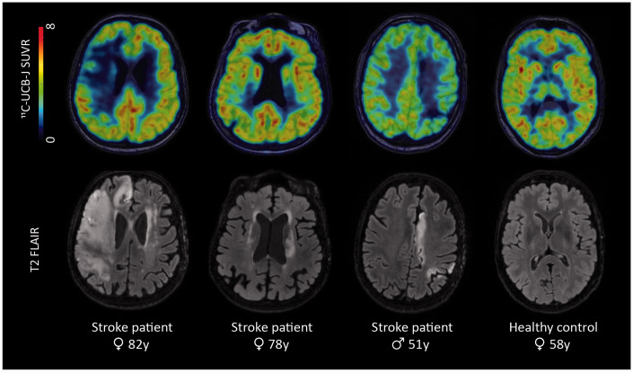
Figure 1.
Representative images of different stroke patients and one healthy control, shown in radiological convention. Top row: 11C-UCB-J SUVR images overlaid on T1-weighted MR images; bottom row: T2 FLAIR images showing the lesions of the patients. Patient 1 was a 82-year old female patient with a large ischemic stroke in the right middle cerebral artery territory. Patient 2 was a 78-year old female patient with a subcortical stroke in the left corona radiata, internal capsule and lentiform nucleus. Patient 3, a 51-year old male, suffered an ischemic stroke in the watershed area of the left hemisphere. The last column shows a 11C-UCB-J SUVR image and T2 FLAIR image of a 58-year-old, female healthy control. SUVR = standardized uptake value ratio.

