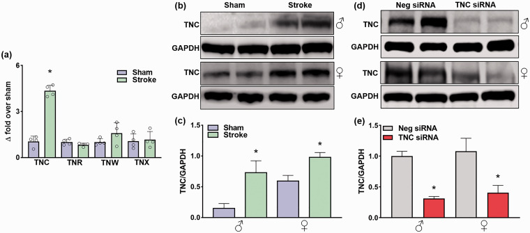
Figure 1.
Focal ischemia significantly increased TNC protein expression, which was inhibited by TNC siRNA treatment. (a) Real-time PCR analysis shows the expression of tenascins at 24 h of reperfusion in the ipsilateral cerebral cortex of adult male mice subjected to 1 h MCAO. Values are mean ± SD; n = 4/group, *p < 0.05 compared with sham by using the Mann-Whitney U test. (b) & (c) TNC protein expression in adult male and female mice at 1 day of reperfusion following transient ischemia. (d) & (e) The TNC siRNA treated cohort showed significant knockdown compared with the non-targeting Neg siRNA treated cohort at 3 days of reperfusion following transient ischemia. The values in the histograms are means ± SD (n = 5/group). *p < 0.05 compared with the respective sham in (b) & (c) and *p < 0.05 compared with the respective Neg siRNA in (d) & (e) by using the Mann-Whitney U test.

