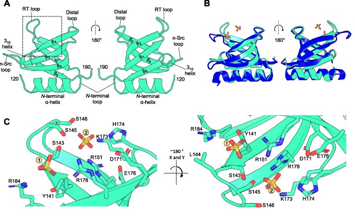
Figure 6.
The BrxR WYL-domain shows ligand binding potential via extensive sidechain coordination. Orthogonal views are shown for each panel as indicated. (A) Close-up of the WYL domain of BrxR. Terminal residues are numbered, and secondary structural elements and loops for this domain are labelled. (B) Structural superposition of BrxREfer with the WYL-domain of PafBC (RMSD 0.662 Å; PDB: 6SJ9) shows clear structural similarity. Differences are observed at the RT loop of BrxREfer, which has moved inwards to bind the two sulphate ions. BrxREfer is shown in cyan and PafBC is shown in blue. (C) A close-up view of the dashed boxed area of (A) shows the hydrogen bond coordination of two sulphate ions bound within the WYL domain of BrxREfer. Interacting sidechains extend from the core β-strands and intervening loops. Nitrogen atoms are shown in blue and oxygen atoms in red. Sulphate ions are shown as yellow (sulphur) and red (oxygen) sticks.

