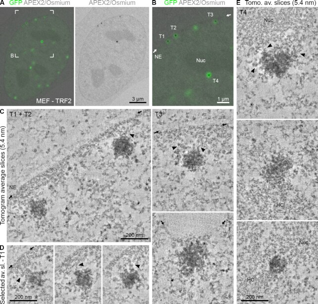
Figure 5.
Telomeres are in direct physical contact with heterochromatin domains. (A) CLEM of MEFs transfected with eGFP–APEX2–TRF2. Left: Overlay image of an optical confocal section (GFP signals, green) and the corresponding TEM image from the physical EM section (APEX2/osmium signals, grey). Right: Corresponding TEM image showing the APEX2/osmium signal only. (B) Magnification of the boxed area in panel (A). NE = nuclear envelope; position indicated by the small arrows. Nuc = nucleolus. (C) Averaged tomographic slices with a thickness of 5.4 nm of the labelled telomeres T1–T3 in panel (B) and one additional example from another cell. Arrowheads point at chromatin contacts between the telomere and non-telomeric heterochromatin. (D) Equivalent to panel (C) showing additional averaged tomographic slices of telomere T1. The position of the NE is only indicated in the first image. (E) Equivalent to panel (C) showing the labelled telomere T4 in panel (B) and additional examples from other cells associated with the nucleolus (Nuc) or other heterochromatin regions (HC).

