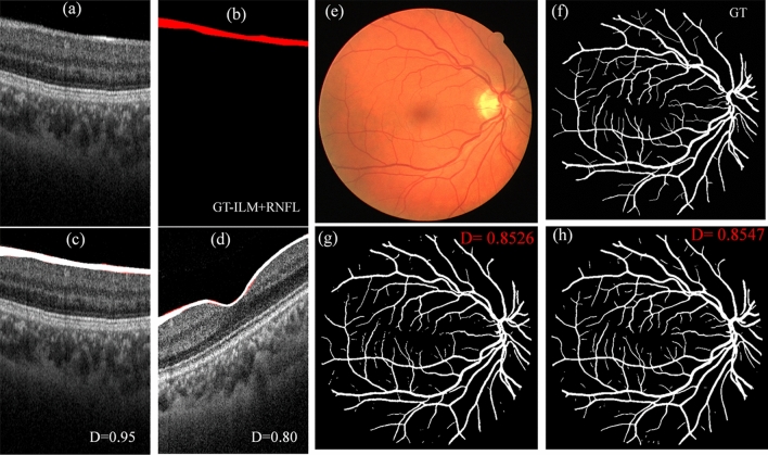
Figure 10.
Application of lightweight DL model in segmentation of other ophthalmic images. (a–d) are the examples of segmentation of ILM + RNFL layer in OCT-B scan, (a) is the original B-scan from the testing dataset which has highest D, (b) is the corresponding manually segmented GT image, (c) is the segmented image overlapped on (a) with GT, and (d) is the segmented image from a testing dataset, which has lowest D. (e–h) are the examples of retinal vessel segmentation on a fundus photo from DRIVE dataset. (e) fundus photo, (f) manual segmentation (from grader-2 in dataset), (g) segmented with Unet_AB, and (h) with LWBNA_Unet model.

