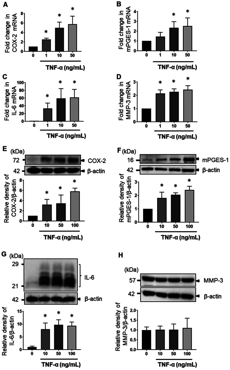
Fig. 2.
MH7A cell response with increasing dose of TNF-α. MH7A cells were cultured in the presence of 1, 10, 50, and 100 ng/mL TNF-α. The mRNA expression levels of COX-2 (A), mPGES-1 (B), IL-6 (C), and MMP-3 (D) were determined by real-time PCR. The expression of COX-2 (E), mPGES-1 (F), IL-6 (G), and MMP-3 (H) in cell lysates was determined by western blot analysis. The data are expressed as the relative protein expression of targets/β-actin. Data are presented as the mean ± SEM of three independent experiments (*P < 0.05 vs. untreated).

