
Figure 9.
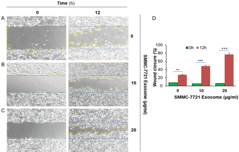
SMMC-7721 liver cancer cells derived exosome induced migration in SMMC-7721 cells. 1×10^6 SMMC-7721 cells were added into 6-well plates for 24 hours until a monolayer formed. Cells were treated with 20 µg/ml of Mitomycin for 3 hours. On the cell surface, we drew a vertical line using 200 µl pipet and washed it with PBS three times. Cells were treated with various concentrations of SMMC-7721 derived exosome (10 µg/ml and 20 µg/ml) at 37°C for 12 hours and were monitored to determine percent closure by microscopy. (A) SMMC-7721 cells were treated with PBS for 0 hour and 12 hours; (B) SMMC-7721 cells were treated with 10 µg/ml of exosomes for 0 hour and 12 hours; (C) SMMC-7721 cells were treated with 20 µg/ml of exosomes for 0 hour and 12 hours, and (D) cells percent closures were obtained after cells were treated with exosomes for 12 hours (23.37%, 48.38%, and 76.6%). Significant difference relative to untreated control are indicated as follows: **P < 0.003, ***P < 0.0006. The images were taken via microscopy: magnification, ×100.