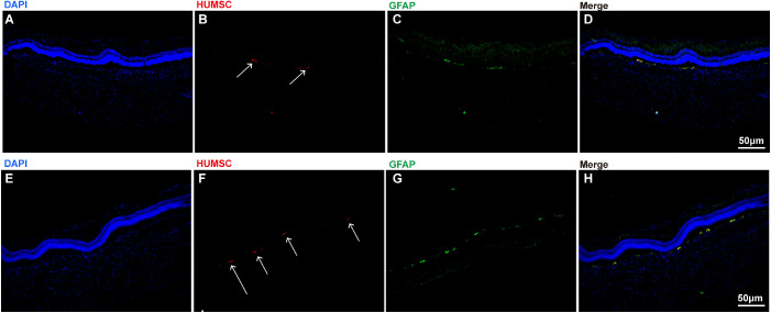
Figure 2.
The retinal distribution and GFAP expression of transplanted HUMSCs in glaucomatous rabbits treated with HUMSC transplantation alone (A, B, C, D) or assisted with UTMD (E, F, G, H). A The structure of retinal tissue stained with DAPI (blue) in rabbits. B HUMSCs labeled with PKH-26 (orange red, directed by arrows). C The expression of GFAP in HUMSCs transplanted into the retina (green, directed by arrows). D The merged image of A to C. E The structure of retinal tissue stained with DAPI (blue) in rabbits. F HUMSCs labeled with PKH-26 (orange red, directed by arrows). G The expression of GFAP in HUMSCs transplanted into the retina (green, directed by arrows). H The merged image of E to G. HUMSCs were directed with arrows. Scale bar, 50 µm.

