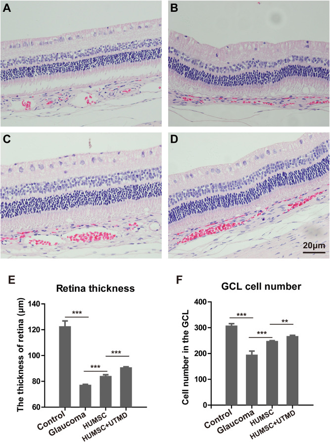
Figure 4.
The structure of retinal tissue with H&E staining in every group of rabbits. (A) The structure of retinal tissue in the control healthy rabbits. (B) The structure of retinal tissue in glaucomatous rabbits without treatment. (C) The structure of retinal tissue in glaucomatous rabbits with HUMSC transplantation. (D) The structure of retinal tissue in glaucomatous rabbits with UTMD-assisted HUMSC transplantation. Scale bar, 20 µm. The thickness of retina (E) and cell number in the GCL (F) in different groups of rabbits. Data were presented as mean values ±SD (error bars). *P < 0.05; **P < 0.01; ***P < 0.001.

