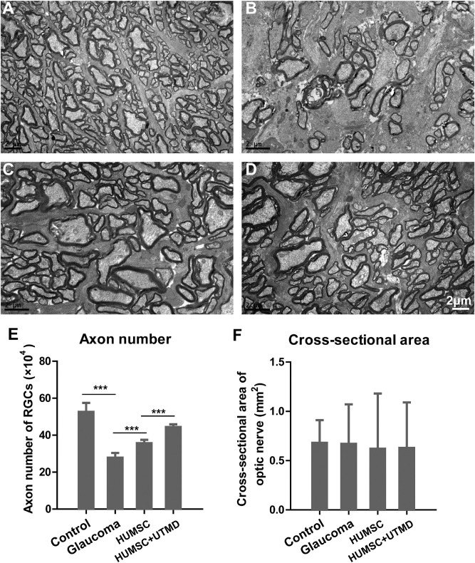
Figure 5.
The structure of optic nerves observed with transmission electron microscope in every group of rabbits. (A) The tissue structure of optic nerve in the control healthy rabbits. (B) The tissue structure of optic nerve in glaucomatous rabbits without treatment. (C) The tissue structure of optic nerve in glaucomatous rabbits with HUMSC transplantation. (D) The tissue structure of optic nerve in glaucomatous rabbits with UTMD-assisted HUMSC transplantation. Scale bar, 2 µm. The axon numbers of RGCs (E) and cross-sectional areas of the optic nerve (F) in different groups of rabbits. Data were presented as mean values ±SD (error bars). *P < 0.05; **P < 0.01; ***P < 0.001.

