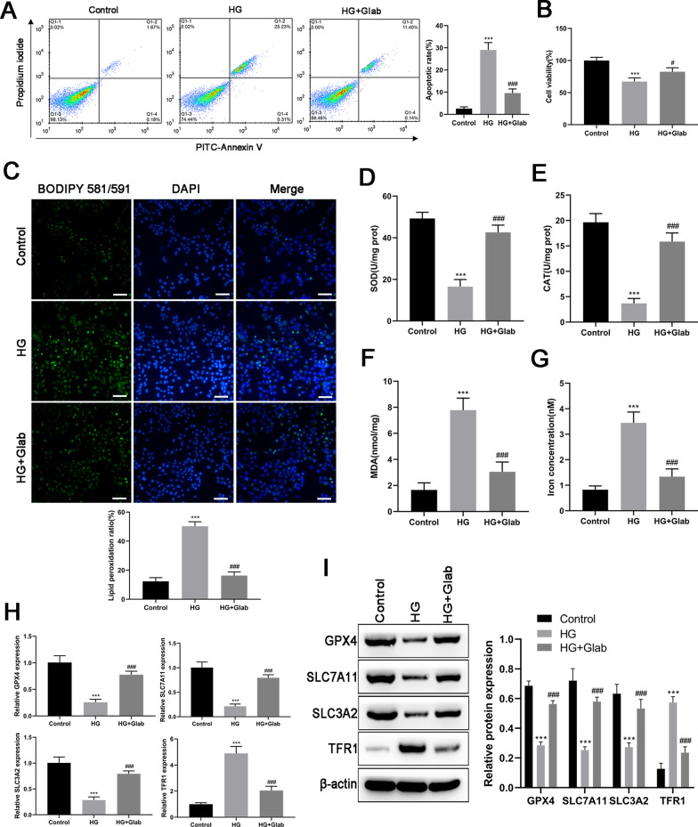
Fig. 6.
Effects of Glab on high glucose (HG) induced oxidative stress and ferroptosis in vitro. The condition of DN in NRK-52E cells was induced by 30 mM glucose. A Cell apoptosis was detected by annexin V-PI double staining. B Cell viability detected by CCK-8 assay. C Lipid peroxidation was determined by the fluorescent probe C11 BODIPY 581/591. Green and blue colors indicate peroxidated lipids and nucleus respectively. The levels of (D) SOD, (E) CAT, (F) MDA, as well as (G) iron concentration were measured by commercial kits. The expression of ferroptosis markers (GPX4, SLC7A11, SLC3A2, and TFR1) at (H) mRNA and (I) protein levels were detected by RT-qPCR and western blot, respectively (***P < 0.005, vs. the control group; #P < 0.05 and ###P < 0.005, vs. the HG group)

