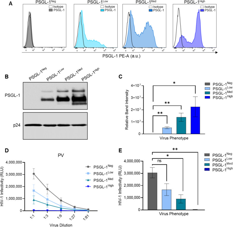
Fig. 2.
Titration of PSGL-1 expression on virus producing cells and the effect on virus infectivity. A Cell surface expression of PSGL-1 on HEK293T cells as detected by flow cytometry 48 h after co-transfection with HIV-1 pseudovirus constructs and increasing amounts of PSGL-1 pDNA (as outlined in Additional file 1: Table S1). Isotype staining is shown with empty histograms, and PSGL-1 staining is shown with filled, coloured histograms (blue or gray). B Semi-quantitative comparisons of virion-incorporated PSGL-1 on pseudovirus stocks via immunoblot analysis. The viral capsid protein p24 was used as loading control to ensure equal loading of total virus lysates across all lanes. This immunoblot is representative of three blots performed showing similar results. C Densitometric quantitation of immunoblot data from B. D Virus infection was tested via normalized viral inputs (displayed as 1:1 in graph), followed by three-fold serial dilutions of viruses. All diluted virus stocks were incubated with TZM-bl reporter cells for 48 h before infectivity was measured using luminescence readout (relative light units; RLU). E Infectivity from the RLU reading with the most concentrated amount of virus (1:1) is shown. For C and E the results of unpaired t tests with Bonferroni correction are shown (*P < 0.05; **P < 0.01). Results show the mean ± SEM of three independent experiments

