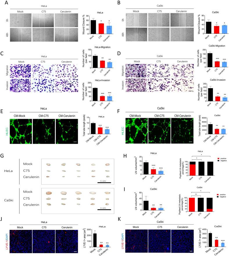
Fig. 7. FASN inhibitors suppress cervical cancer cell migration, invasion, lymphangiogenesis in vitro, and lymph node metastasis in vivo.
A, B The wound healing assay results in the mock-, C75-, and Cerulenin-treated HeLa and CaSki cells. Bar = 200 µm. C, D Transwell migration and invasion results in the same settings as A, B. Bar=50 µm. E, F Assessment of lymphangiogenesis by tube formation assays in the CM-mock (mock vehicle-treated tumor culture medium) and CM-C75 or CM-Cerulenin (C75- or Cerulenin-treated tumor culture medium) groups and the total tube length. Bar = 100 µm. G A gross popliteal lymph node specimen is shown. Bar=1 cm. H, I LN volume and metastasis percentage were quantified in the mock, C75, and Cerulenin groups using both cell lines injected into the CC lymph node mouse model. J, K LYVE-1 immunofluorescence staining showed the lymphatic vascular density in the same setting groups as A, B. Bar=25 µm. *P < 0.05; **P < 0.01; ***P < 0.001.

