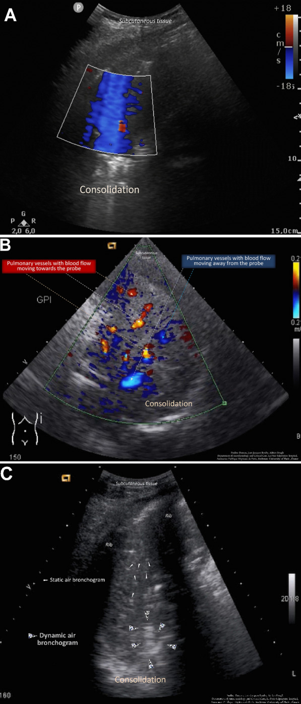
Fig. 1.

A Color Doppler lung ultrasound in the consolidated left lower lobe in a 54-year-old patient without pneumonia on venoarterial extracorporeal membrane oxygenation. The extracorporeal membrane oxygenation was initiated 2 days before and pneumonia was ruled out by a bronchoalveolar lavage retrieving 2.102 orophayngeal flora. Color signals are diffuse and changing resulting from interferences caused by respiratory movements and heart beats. Corresponding to Video S1A (Additional File 1). B Color Doppler intrapulmonary flow detected in the consolidated right lower lobe of a 43-year-old patient with pneumonia on venoarterial extracorporeal membrane oxygenation. The extracorporeal membrane oxygenation was initiated 12 days before for a cardiogenic shock and pneumonia was confirmed by a bronchoalveolar lavage retrieving 104 Escherichia coli. The blood flow in a vessel is seen as a color signal persisting in the same location during the respiratory cycle with a tubular, curvilinear, or branching distribution on real-time images. When blood flow signals are detected, pulse-wave Doppler can identify their spectral waveform.19 Corresponding to Video S1B (Additional File 2). C Dynamic air bronchogram in a 79-year-old patient with pneumonia on venoarterial extracorporeal membrane oxygenation. The extracorporeal membrane oxygenation was initiated 5 days before for a cardiogenic shock and pneumonia was confirmed by a bronchoalveolar lavage retrieving 6.106 Hafnia alvei. Corresponding to Video S1C (Additional File 3)
