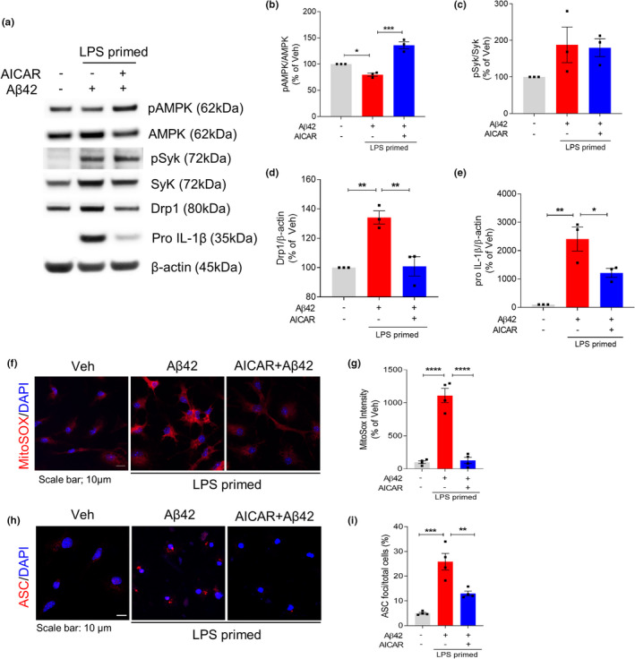
FIGURE 4.

AMPK activation restores Aβ42‐induced mitochondrial hyperfission and inflammasome activation in LPS‐primed microglia. LPS‐primed microglia were pretreated with AICAR (1 mM) for 1 h and then treated with Aβ42 for 24 h. (a–e) Representative immunoblot (a) and densitometric quantifications (b–e) of the protein levels of pAMPK, AMPK, pSyk, Syk, Drp1, pro‐IL‐1β, and β‐actin in total cell lysate. (f–g) MitoSOX (red) and DAPI (blue) staining and quantification of MitoSOX fluorescence intensity. Scale bar: 10 μm. (h) Immunostaining of ASC (red) and DAPI (blue). Scale bar: 10 μm. (i) Percentages of microglia containing ASC foci were counted. Data are shown as mean ± SEM from at least three independent experiments and are analyzed by one‐way ANOVA and Tukey's multiple comparisons test. *p < 0.05, **p < 0.01, ***p < 0.001, and ****p < 0.0001
