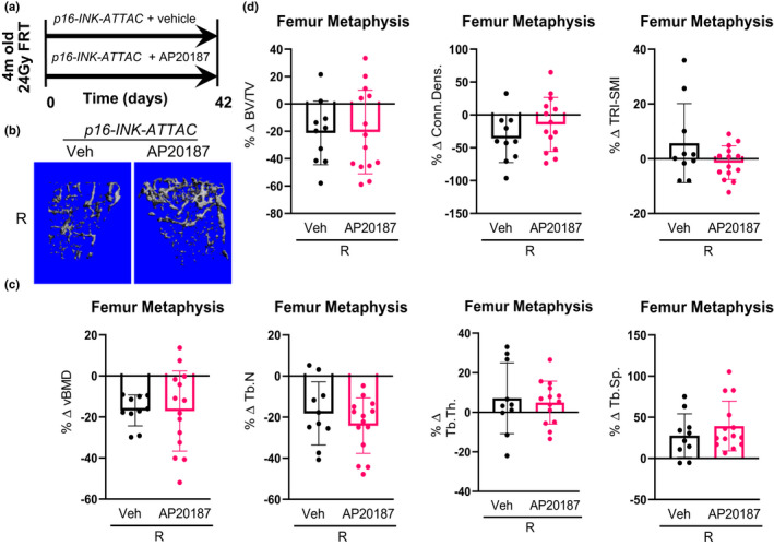
FIGURE 5.

Targeted clearance of p16Ink4a ‐expressing cells has no effect on radiation‐induced bone damage. (a) Schematic of the experimental design. The right legs of p16‐INK‐ATTAC mice were radiated (24 Gy) near the femoral metaphysis (5 mm above the growth plate), while the left legs served as control. The animals were assigned to vehicle‐ (n = 10) or AP20187‐ (n = 14) treated groups receiving doses twice weekly for 6 weeks. R‐ and NR‐femurs at 42 days postradiation were collected for μCT analysis. (b) Images are 3‐dimensional representations of the bone architecture generated by ex vivo μCT scans. All quantifications are done as percentage difference between the R leg vs. the NR control leg and comparisons are between vehicle‐ and AP20187‐treated radiated bones. (c) Percentage change of volumetric bone mineral density, and (d) bone architecture parameters: bone volume fraction (BV/TV), connectivity density (Conn.Dens.), structural modal index (SMI), trabecular number (Tb.N), trabecular thickness (Tb.Th.) and trabecular separation (Tb.Sp.). Statistical comparison was done using a two‐tailed unpaired t test between the Veh‐R and AP20187‐R bones
